Cell Fusion
Cell Fusion
The extensive studies carried out to localize the genes involved in XP have only been successful recently. Indeed, traditional strategies such as the use of interspecies proliferating hybrids have been unsuccessful for NER genes mapping.
As former techniques didn’t work, scientists used cell fusion, a natural process that can be reproduced artificially. The cell fusion is a process in which two cells are merged to form 1 new cell with twice as much genetic information.
In the case of XP, scientists used cell fusion to find how many genes were involved in the disease. First of all, they took XP cells from two patients, and merged them together. In certain cases, the new cell was not an XP cell. They concluded that the genes involved for both patients were not the same. By doing this with many different patients, they found out that 7 different genes were involved.
With another experiment, they discovered that all the genes were independent, and even that they were all located on different chromosomes. This experiment is described in the scheme below.
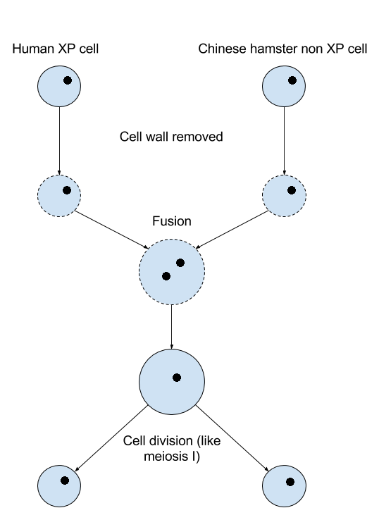
Once that done many times, they exposed all the final cells to sunlight (thus UV), in order to kill all XP cells. What they found out is that if you randomly take one survivor cell (non XP cell, as it survived UV), and you look at its chromosomes 2, 3, 9, 11, 13, 16, 19 (more precisely where they come from), they would never be all coming from the human. All combinaison were possible, except the ones that had all of these chromosomes from human, which means that these cells were necessarily killed by UV, (=XP cells). That’s how they concluded that the 7 genes that, once mutated, caused XP were on 7 different chromosomes.
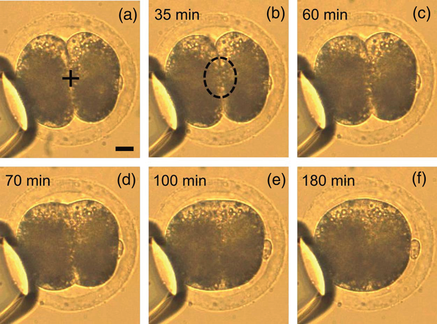 |
| Microscopic observation of Cell Fusion |
Here’s an example :
Let’s take two examples of merged cells, before exposition to sunlight.
Bold numbers : chromosomes involved in XP.
The first cell contains chromosomes :
From patient: 1, 4, 6, 9,11,12, 15, 17, 18, 19, X
From hamster : 2, 3, 5, 7, 8, 10, 13, 14, 16, 20, 21
If the patient’s mutation is on chromosome 9,11, or 19, the cell will die to UV, else it will survive.
The second one contains chromosomes :
From patient: 2, 3, 4, 9, 11, 12, 13, 14, 15, 16, 17, 19, X
From hamster : 1, 5, 6, 7, 8, 10, 18, 20, 21
Whatever chromosome the mutation is on, the cell will die to UV anyway.
If all chromosomes in bold come from human, wherever the other chromosomes come from, the cell will die to UV. It means that a survivor cell cannot contain all bold chromosomes from human.
Producing mutant of interest
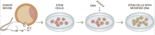
When they knew which genes were involved in xeroderma pigmentosum, scientists used gene targeting in mouse embryonic cells to produce mutants of interest. Indeed, they wanted to discover what implied a mutation in the genes they found, and for obvious ethic reasons, they could not do some experiments on XP human patients. Furthermore, those patients were really rare and died quickly so they were very difficult to study.
Gene targeting consists in introducing a genetic change into the mouse ES cells. Those containing the desired modification are selected, enriched and then, microinjected into mouse blastocysts, which are injected into foster mothers. The mice born from these embryos are chimeric. They are able to transmit the mutation to their progeny, so they are crossed, again and again, until finding homozygous mice that are interesting to study XP. Indeed, they could exhibit characteristic features that mimic XP patients.

Now, you should wonder what they did to those mutants and how those experiments were useful to understand the mechanisms involved in xeroderma pigmentosum for humans. We will not explain here all the experiments that were done, but we will try to show you some of the most important.
XPA-Deficient animals

The mutants of 8-10 weeks were, for exemple, irradiated on the back with UV at a dose of 5 J/cm2 three times weekly for 10 weeks. More than 30% of the irradiated mice developed skin tumors, whereas non-irradiated ones did not develop it spontaneously.
XPA mice also appeared to develop internal tumors with a much higher frequency and a shorter latency than normal mice when they are exposed to different carcinogens.
The information that those experiments give is in agreement with what scientists thought about the illness: xeroderma pigmentosum, when XPA is mutated, results in predisposition to skin cancers when patients are exposed to sunlights.
Other mutants
Scientists did quite similar experiments on XPB,C,D,E,F and G deficient mice, but they also tested other things like, for example, neurological problems for XPG mutants.
Indeed, as homozygous mice for mutated XPG developed a diminished level of activity and progressive ataxia were observed, suggesting a developmental retardation of the central nervous system and a progressive neuronal dysfunction. Scientists decided to weight the brain of those mutants and of the wild type mice at day 19. Here are the results:

Conclusion on those experiments
Scientist recently made lots of discoveries on XP. The interesting mouse model they developed have made them understand the complex mechanisms of that illness. We now know exactly what are the consequences of each possible mutation causing XP. However, the goal of all of this is to be able to find a cure for human patients, and we are still far away from it.
No comments:
Post a Comment